
Fetal echocardiography in Vijayawada is a highly specialized ultrasound examination designed to evaluate the structure and function of a baby’s heart while still in the womb. Unlike routine pregnancy scans, this detailed imaging technique focuses exclusively on the fetal cardiovascular system, allowing doctors to identify congenital heart defects, rhythm abnormalities, and blood flow disturbances at an early stage of development. By offering a precise and non-invasive view of the fetal heart, fetal echocardiography plays a pivotal role in ensuring timely diagnosis, counseling, and treatment planning for expectant parents.
UNDERSTANDING FETAL ECHOCARDIOGRAPHY
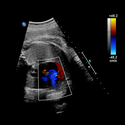
Fetal echocardiography or Fetal heart scan in Vijayawada uses high-frequency sound waves to create real-time images of the baby’s heart chambers, valves, major blood vessels, and rhythm patterns. It is usually performed between the 18th and 24th weeks of pregnancy, when the fetal heart is sufficiently developed for accurate assessment. However, in high-risk pregnancies, it may be conducted earlier or repeated later for closer monitoring.
This investigation is not painful, does not involve radiation, and is completely safe for both the mother and the baby. The procedure provides dynamic images that allow specialists to observe how the fetal heart beats, pumps blood, and coordinates electrical activity.
WHEN IS FETAL ECHOCARDIOGRAPHY RECOMMENDED?
While many pregnancies progress normally, certain conditions increase the risk of fetal heart abnormalities. Congenital heart defect scan in Vijayawada is commonly advised in the following situations:
- Family history of congenital heart disease
- Diabetes, lupus, or other chronic illnesses in the mother
- Exposure to certain medications or infections during pregnancy
- Abnormal findings on routine ultrasound
- Suspected chromosomal or genetic disorders
- Multiple pregnancies or assisted reproductive techniques
Early referral for this test ensures that any potential cardiac concern is detected long before birth, enabling well-planned medical care.
WHAT CONDITIONS CAN BE DETECTED?
Fetal echocardiography is capable of identifying a wide spectrum of heart-related conditions, including:
- Structural defects such as holes in the heart walls or malformed valves
- Abnormal heart rhythms like fetal arrhythmias
- Major vessel anomalies affecting blood flow to and from the heart
- Cardiomyopathies involving the heart muscle
- Complex congenital heart diseases requiring surgical or medical intervention after birth
The clarity and precision of this scan allow specialists to differentiate minor variations from life-threatening conditions.
THE PROCEDURE: WHAT TO EXPECT
During a fetal echocardiography session, the mother lies comfortably while a specialized transducer is moved gently over the abdomen. A gel is applied to ensure smooth contact and better image transmission. The process typically takes between 30 and 60 minutes, depending on fetal position and the complexity of the evaluation.
The cardiologist carefully studies multiple views of the heart, assessing:
- Chamber size and symmetry
- Valve movement and integrity
- Direction and speed of blood flow
- Heart rate and rhythm
Results are usually discussed soon after the scan, with detailed explanations provided to the parents.
BENEFITS OF EARLY CARDIAC DIAGNOSIS
Identifying heart abnormalities before birth offers significant clinical and emotional advantages. These include:
- Timely medical planning for delivery at a specialized center
- Immediate neonatal care for babies with critical heart defects
- Parental counseling to understand prognosis and treatment options
- Reduced complications through proactive intervention
In many cases, prenatal detection dramatically improves survival rates and long-term outcomes.
MULTIDISCIPLINARY CARE AND FOLLOW-UP
A fetal echocardiography diagnosis often leads to a collaborative care approach involving obstetricians, fetal medicine specialists, pediatric cardiologists, and neonatologists. This team works together to monitor the pregnancy, prepare for delivery, and arrange any necessary postnatal procedures.
Follow-up Targeted anomaly scan in Vijayawada may be scheduled to observe how the condition evolves as the baby grows. After birth, newborn echocardiography confirms the findings and guides further treatment, which may include medication, catheter-based procedures, or surgery.
A VITAL STEP TOWARD A HEALTHY FUTURE
Fetal echocardiography represents a powerful tool in modern prenatal medicine. By unveiling the intricate workings of a baby’s heart before birth, it offers families clarity, preparedness, and hope. With early detection and expert medical guidance at the Best Fetal Medicine Center in Vijayawada, many babies born with heart conditions go on to lead healthy, fulfilling lives. This advanced screening is not just a diagnostic test—it is a crucial step toward safeguarding the heart of a new life.
At Neo Life Fetal Scan Centre , under the expert leadership of Dr. V. Prashanthi, fetal echocardiography is performed with the highest standards of precision, compassion, and clinical excellence. Combining advanced imaging technology with meticulous interpretation, we are committed to the early and accurate detection of fetal cardiac conditions, enabling timely guidance, informed decision-making, and the best possible outcomes for both mother and baby.